Composite Illustration of A Cell And Its Cytoplasmic Organelles (DR HC 0001)
Different Types of Epithelia In Selected Organs (DR HC 0002)
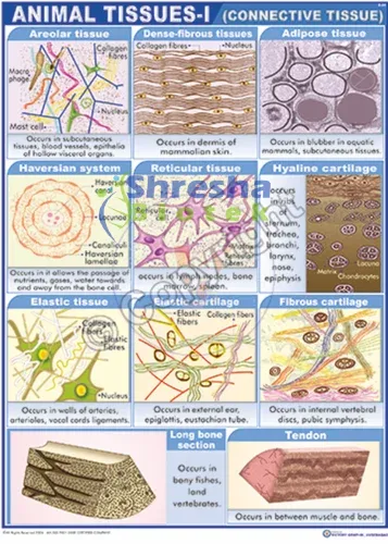
Composite Illustration of Loose Connective Tissue with Its Predominant Cells and Fibers (DR HC 0003)
Endochondral Ossification, Illustrating the Progressive Stages of Bone Formation (from Cartilage Model to Bone) and Including the Histology of A Section of Formed Bone (DR HC 0004)
Differentiation of A Pluripotential Hemopoietic Stem Into the Myeloid Stem Cell Line and Lymphoid Stem Cell Line During Hemopoiesis (DR HC 0005)
Microscopic Illustrations of the Three Types of Muscles: Skeletal, Cardiac, and Smooth (DR HC 0006)
The Central Nervous System Composed of the Brain and Spinal Cord (DR HC 0007)
The Peripheral Nervous System: Cranial and Spinal Nerves (DR HC 0008)
Comparison of Muscular Artery, Large Vein, and Three Types of Capillaries (DR HC 0009)
Location and Distribution of the Lymphoid Organs and Lymphatic Channels in the Body (DR HC 0010)
Comparison Between Thin Skin in the Arm and Thick Skin in the Palm (DR HC 0011)
Simple Squamous Epithelium: Peritoneal Mesothelium Surrounding Small Intestine (DR HC 0012)
Simple Columnar Epithelium: Stomach Surface (DR HC 0013)
Simple Columnar Epithelium on Villi in Small Intestine (DR HC 0014)
Pseudostratified Columnar Ciliated Epithelium: Respiratory Passages (Trachea) (DR HC 0015)
Transitional Epithelium: Bladder (Contracted) (DR HC 0016)
Stratified Squamous Nonkeratinized Epithelium: Esophagus & Keratinized Epithelium: Palm of the Hand (DR HC 0017)
Stratified Cuboidal Epithelium: Excretory Duct In Salivary Gland (DR HC 0018)
Unbranched Simple Tubular Exocrine Glands: Intestinal Glands (DR HC 0019)
Coiled Tubular Exocrine Glands: Sweat Glands (DR HC 0020)
Loose Connective Tissue (DR HC 0021)
Individual Cells of Connective Tissue (DR HC 0022)
Loose Connective Tissue & Dense Irregular and Loose Irregular Connective Tissue (Elastin Stain) (DR HC 0023)
Ovary: Dog (Panoramic View) (DR HC 0024)
Ovary: Ovarian Cortex and Primary and Primordial Follicles (DR HC 0025)
Uterine Tube: Ampulla (Panoramic View, Transverse Section) (DR HC 0026)
Uterus: Proliferative (Follicular) Phase (DR HC 0027)
Vagina (Longitudinal Section) (DR HC 0028)
Vagina: Surface Epithelium (DR HC 0029)
Placenta at 5 Weeks (Panoramic View) (DR HC 0030)
Inactive Mammary Gland (DR HC 0031)
Mammary Gland During Proliferation and Early Pregnancy (DR HC 0032)
Eyelid (Sagittal Section) (DR HC 0033)
Lacrimal Gland & Cornea (Transverse Section) (DR HC 0034)
Whole Eye (Sagittal Section) & Retina, Choroid, and Sclera (Panoramic View) (DR HC 0035)
Inner Ear: Cochlear Duct (Scala Media) (DR HC 0036)
Format: Chart with lines of letters, starting with a large single letter at the top
Letter Type: Sans-serif, standardized optotypes (not standard alphabet fonts)
Distance: To be used at a standard distance of 6 meters (20 feet)
Visual Acuity Markings: Each line marked with corresponding Snellen fraction (e.g., 6/6, 6/9, 6/12, 6/18, 6/24, 6/36, 6/60, etc.)
Top Letter Size: Large enough to be seen by a person with 6/60 vision at 6 meters
Bottom Line Size: Small enough to correspond to 6/6 normal vision at 6 meters
Material: High-quality, non-reflective board or laminated plastic, or digitally printed on matte surface
Language: Usually in English letters, also be available in regional scripts or E-charts for non-literate patients
Dimensions: around 22 x 56 cm (vary slightly depending on format and design)
©2025-26. All Rights Reserved. Shresha Biortek PVT LTD